
Single-step Staining Reaction—Time-saving and Labor-efficient.
High Sensitivity and Strong Specificity.
Suitable for Detection of Late-stage Apoptosis.
Cat. No. :
YBS2607-50TBrand :
YanbiotechProduct Origin :
ChinaVolume :
50TI. Product Introduction
During apoptosis, chromosomal DNA undergoes a progressive, stepwise fragmentation. Initially, endogenous nucleases cleave chromosomal DNA into large fragments of 50–300 kb. Subsequently, approximately 30% of the chromosomal DNA is randomly cleaved at internucleosomal sites by Ca²⁺- and Mg²⁺-dependent endonucleases, generating 180–200 bp multimers of nucleosomal DNA. Thus, in the late stages of apoptosis, DNA is degraded into 180–200 bp fragments, exposing numerous 3'-OH termini on the fragmented genomic DNA.
Terminal Deoxynucleotidyl Transferase (TdT) is a template-independent DNA polymerase that catalyzes the incorporation of deoxynucleotides onto the 3'-OH ends of DNA breaks. The TUNEL (TdT-mediated dUTP Nick End Labeling) assay leverages this enzyme to detect nuclear DNA fragmentation in cells during late apoptosis. The principle involves the TdT-mediated incorporation of biotin-labeled dUTP (Biotin-dUTP) at the 3'-OH ends of fragmented genomic DNA. Subsequently, horseradish peroxidase (HRP)-labeled streptavidin (Streptavidin-HRP, SA-HRP) binds to the biotin-labeled DNA ends. Finally, the HRP substrate DAB is applied, producing a chromogenic reaction that stains apoptotic nuclei brown-yellow, enabling detection under a standard light microscope.
This versatile kit is suitable for detecting apoptosis in various sample types, including paraffin-embedded tissue sections, frozen tissue sections, cell smears, and cytospin preparations.
II. Product Information:
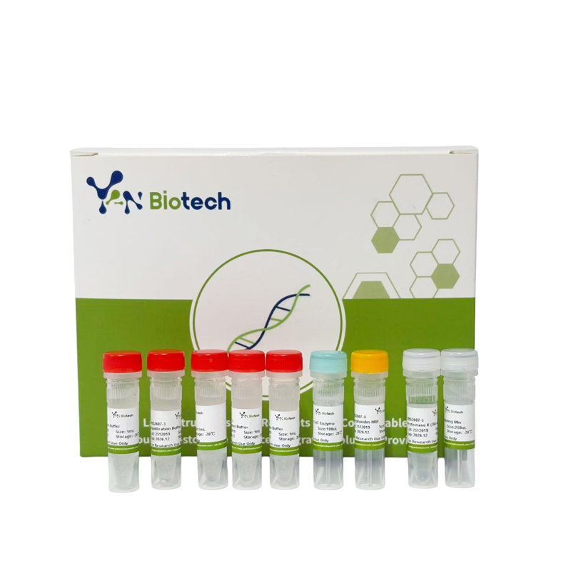
| Component Number | Component | YBS2607-50T | YBS2607-100T |
| 50T | 100T | ||
| YBS2607-1 | Recombinant TdT Enzyme | 100 µL | 2 × 100 µL |
| YBS2607-2 | Biotin-dUTP Labeling Mix | 250 μL | 2 × 250 µL |
| YBS2607-3 | Equilibration Buffer | 5 × 1 mL | 10 × 1 mL |
| YBS2607-4 | Streptavidin-HRP | 25 µL | 2 × 25 µL |
| YBS2607-5 | Proteinase K (200 µg/mL) | 1 mL | 2 × 1 mL |
III. Storage Condition:
Transport: Wet ice.
Storage: Store at –20°C. Shelf life: 12 months.
IV. Procedure (Please read the precautions before starting the experiment):
I. Sample Preparation
A. Paraffin-Embedded Tissue Sections
1. Deparaffinize and rehydrate sections:
2. Rinse gently with PBS and remove excess liquid around the sample. Use an immunohistochemistry pen to draw a circle 2–3 mm from the tissue edge to facilitate permeabilization and equilibration. Keep samples moist at all times; place in a humidified chamber.
3. Prepare Proteinase K working solution: Dilute Proteinase K stock (200 µg/mL) 1:9 with PBS to a final concentration of 20 μg/mL.
4. Apply 100 μL of Proteinase K working solution to each sample, ensuring complete coverage. Incubate at 37°C for 20 min.
(Note: Proteinase K treatment enhances reagent penetration. Both under- and over-treatment may affect labeling efficiency; optimization may be required.)
5. Wash samples 3 times with PBS, 5 min each. Ensure complete removal of Proteinase K, as residual enzyme may interfere with subsequent labeling. Keep samples moist in a humidified chamber.
6. (Optional) Remove excess liquid, apply permeabilization buffer to cover the tissue, and incubate at room temperature for 20 min. Wash 3 times with PBS, 5 min each. Keep samples moist.
7. Remove excess liquid, apply 3% H₂O₂ (prepared in PBS) to cover the tissue, and incubate at room temperature for 20 min to quench endogenous peroxidase activity. Prolonged incubation may cause H₂O₂-induced DNA damage, leading to false positives. Wash 3 times with PBS, 5 min each. Keep samples moist.
B. Frozen Tissue Sections
1. Fix slides in 4% paraformaldehyde (in PBS) at room temperature for 10–15 min.
2. Air-dry slides in a fume hood.
3. Rinse slides in distilled water or PBS to remove residual fixative.
4. Draw a circle around the tissue with an immunohistochemistry pen. Keep samples moist at all times.
5. Prepare Proteinase K working solution as described in A3.
6. Apply 100 μL of Proteinase K working solution to each sample and incubate at room temperature for 10 min.
7. Wash 2–3 times with PBS to remove Proteinase K. Keep samples moist.
8. (Optional) Permeabilize as described in A6.
9. Quench endogenous peroxidase with 3% H₂O₂ as described in A7.
C. Cell Smears
1. Resuspend cells in PBS at approximately 2 × 10⁷ cells/mL. Pipette 50–100 μL onto adhesive slides and gently spread with a clean coverslip.
2. Fix slides in 4% paraformaldehyde (in PBS) at 4°C for 25 min.
3. Wash slides in PBS at room temperature for 5 min; repeat once.
4. Permeabilize in permeabilization buffer at room temperature for 5 min.
*(Note: Alternatively, treat with 2–20 μg/mL Proteinase K at 37°C for ~10 min, depending on cell type and adhesion.)*
5. Wash 2–3 times in PBS. Apply 3% H₂O₂ (in PBS) and incubate at room temperature for 20 min.
6. Remove excess liquid, wash 3 times with PBS, 5 min each. Carefully aspirate liquid around the sample. Keep samples moist.
D. Cytospin Preparations
1. Culture adherent cells in Lab-Tek Chamber Slides. After apoptosis induction, rinse gently twice with PBS.
2. Add sufficient 4% paraformaldehyde (in PBS) to each chamber and fix at room temperature for 20 min.
3. Remove fixative and wash 3 times with PBS, 5 min each.
4. Permeabilize in permeabilization buffer at room temperature for 5 min.
(Note: Alternatively, use Proteinase K as described above.)
5. Wash 2–3 times in PBS. Add 3% H₂O₂ (in PBS) and incubate at room temperature for 20 min.
6. Remove liquid, wash 3 times with PBS, 5 min each. Carefully dry area around sample with filter paper. Keep samples moist.
V. Labeling and Detection:
1. Equilibration: Apply 50 μL of Equilibration Buffer to each sample, ensuring complete coverage. Incubate at room temperature for 10 min.
2. Preparation of Labeling Mix: Thaw Biotin-dUTP Labeling Mix and Equilibration Buffer on ice. Prepare TdT incubation buffer as follows (per sample):
| Component | Volume |
| Recombinant TdT Enzyme | 2 µL |
| Biotin-dUTP Labeling Mix | 5 µL |
| Equilibration Buffer | 50 µL |
Adjust volumes proportionally based on sample size.
3. Negative Control: Prepare a control TdT incubation buffer without Recombinant TdT Enzyme, substituting with ddH₂O.
4. Labeling: Remove equilibration buffer completely. Add 57 μL of TdT incubation buffer to each sample. Incubate at 37°C for 1 h in a humidified, light-protected chamber. Avoid drying.
5. Wash: Rinse slides immediately with PBS, 4 times, 5 min each.
6. Gently blot PBS around the sample with filter paper.
7. Streptavidin-HRP Reaction: Remove excess liquid and apply 100 µL of diluted Streptavidin-HRP (dilute Streptavidin-HRP 1:200–1:500 in TBST) to each sample. Incubate at 37°C for 30 min.
8. Wash: Wash 3 times with PBS, 5 min each.
9. DAB Chromogenic Reaction: Prepare DAB working solution immediately before use (recommended: G1212). Apply 50–100 µL per sample and monitor color development under a microscope. Once positive staining appears, immediately transfer slides to a humidified chamber and rinse with distilled water to terminate the reaction.
10. Hematoxylin Counterstaining:
11. Dehydration and Clearing:
12. Mounting: Mount coverslips with neutral balsam. Air-dry or dry in a 60°C oven.
13. Microscopy: Examine under a light microscope. Apoptotic nuclei appear brown-yellow; normal nuclei appear blue.
For Research Use Only.

Stay Connected With Us


Copyright @ Wuhan Yanbiotech Co., Ltd. All Rights Reserved.
 Network Supported Xml / Privacy Policy
 English
 English
 Русский
 Español